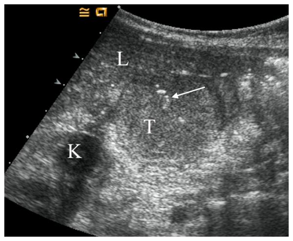
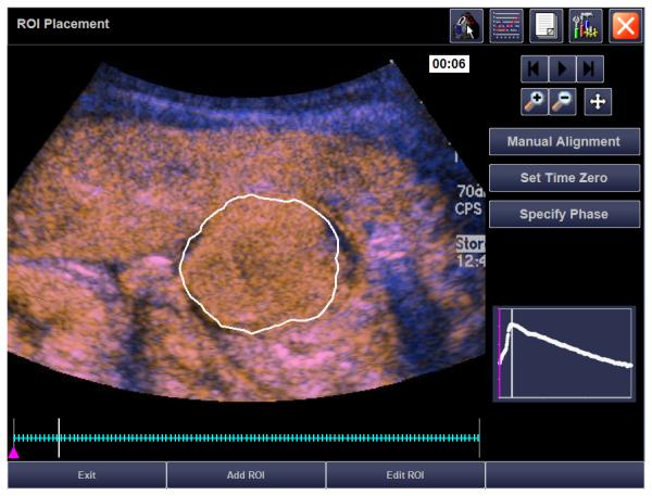
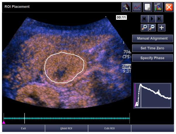
Figure2.


21 month old girl with recurrent rhabdoid tumor. A) Transverse grey-scale sonogram shows a peritoneal tumor (T) located posterior to the liver (L) and medial to the right kidney (K). Tumoral calcification (arrow) and adjacent organs were used as landmarks for transducer placement. B) Baseline transverse CEUS image with ROI inside tumor margins, obtained at PE of 33.5 dB. C) Day 7 after initiation of therapy, transverse CEUS image obtained at PE of 30.6 dB giving a 8.7% reduction compared to baseline. This subject progressed before the end of course 1, at 22 days.
