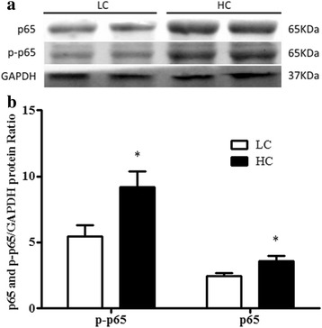
Fig. 3.

a The western blotting assay of NF-κB (p65) and phosphorylated-p65 protein in the uterus. The NF-κB contents in uterus were evaluated through western blot of the low-concentrate (LC) group and high-concentrate (HC) group. b The quantities of proteins are measured as arbitrary units relative to GAPDH; fold alterations in NF-κB (p65) and phosphorylated NF-κB (p-p65). The data were expressed as the mean ± SEM., asterisks indicate the differences between the high-concentrate (HC) group compared to the low-concentrate (LC) group (** p < 0.01)
