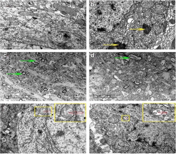
Fig. 4.

Alterations in hippocampus ultrastructure was examined by transmission electron microscope. Autophagosomes (red arrow) existed in both control and diabetic mice were no significantly decrease in diabetes mice. Diabetes mice have more lipofuscin (yellow arrow) and fewer myelinated nerve fibers (green arrow) in the hippocampus. a, c, e were control groups and (b, d, f) were diabetes groups
