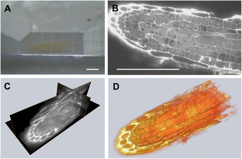
Fig. 2.

Fluorescence light microscopy on serial sections from Arabidopsis roots. Arrays produced from Arabidopsis roots embedded in HM20 (a) were stained with propidium iodide and imaged in a standard wide field fluorescence microscope (b). 3D reconstructions from a stack of 200 sections were visualized in Amira as orthoslices (c) or by volume rendering (d). Scale bars: 100 μm
