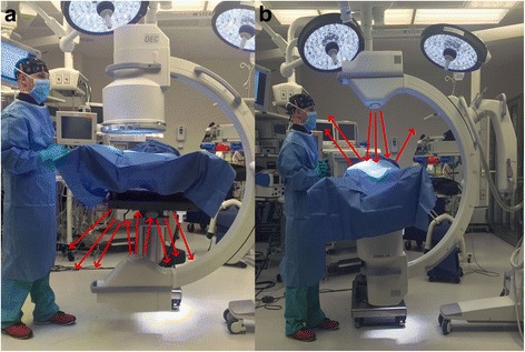
Fig. 3.

a shows a setup with the x-ray tube on the bottom. The red arrows represent radiation beams that scatter after they deflect off of the object being imaged. With the x-ray tube on the bottom most of the scattered (deflected) radiation is towards the legs and feet of the surgeon. b shows a setup with the x-ray tube on the top. Here the scattered radiation is towards the head and neck region of the surgeon
