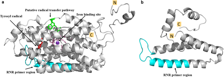
Figure 3. Predicted 3-D structures of nrdBL1 (long form) and nrdBS1 (short form) of ribonucleotide reductase β-subunit.
(a) nrdBL and (b) nrdBS. The iron binding residues in pink centered by a purple dot (binding site), the tyrosyl radical in red, the putative radical transfer pathway in green. The regions targeted by primer set RNRf/RNRr were highlight in cyan. All conserved residues and model was generated using Phyre server29. The final refinement of all 3-D structure figures were made using the Pymol Molecular Graphics System (v1.7.6).

