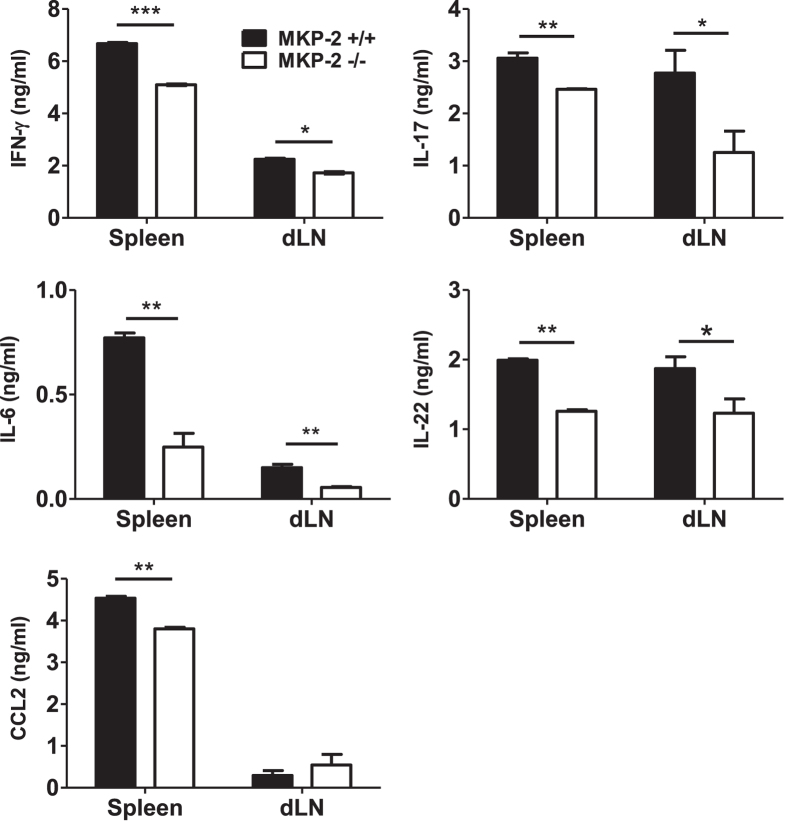
Figure 3. MKP-2−/− mice display a reduced immune response during EAE development compared to MKP-2+/+ mice.
Spleens and dLNs were harvested at EAE peak and disrupted to form individual cell suspensions. Cells were stimulated with or without MOG35-55 for 72 hours and supernatants collected for analysis of cytokine expression by ELISA. Graphs show mean ± SEM of three independent experiments, n = 12 per group *P < 0.05; **P < 0.01, ***P < 0.001.

