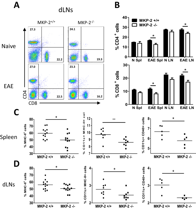
Figure 4. Altered phenotypte of immune cells in naïve and EAE MKP-2+/+ and MKP-2−/− mice lymphoid organs.
(A and B) Cells from naïve and EAE spleen and dLNs were stained for CD4 and CD8 and analysed by flow cytometry. (A) FACS plots of dLN tissue cells are representative of at least 10 mice per EAE group (MKP-2+/+ and MKP-2−/− mice) from three independent experiments. (B) Bar graphs show combined data from three independent experiments with n = 10 in each group, N refers to naïve, and Spl refers to spleen in the x-axis labelling, bars represent mean ± SEM. (C and D) Expression of MHC-II, CD11c and CD40 by MKP-2+/+ and MKP-2−/− immune cells in EAE spleen (C) and dLNs (D). Spleen and dLNs were harvested at EAE peak and disrupted to form individual cell suspensions. Cells were then stained and analysed for expression of MHC-II, or expression of both CD11c and MHC-II molecules, or both CD11c and CD40 by flow cytometry. Graphs show combined data from three independent experiments, each symbol represents an individual mouse and bar indicates the average value of expression in each group. *P < 0.05; **P < 0.01.

