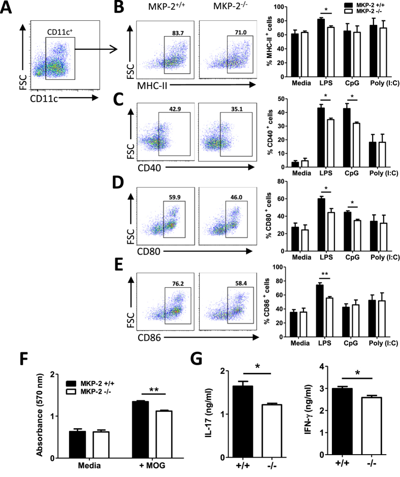
Figure 6. BMDCs of MKP −/− mice exhibit reduced expression of MHC-II and costimulatory molecules.
BMDCs were generated from the culture of bone marrow from MKP-2+/+ and MKP-2−/− mice. Cells were stimulated with or without LPS (100ng/ml), CpG (1 μM) or Poly(I:C) (20 μg/ml) for 24 hours and CD11c, MHC-II, CD40, CD80 and CD86 expression analysed by flow cytometry. FACS plots are representative of three independent experiments of BMDCs activated by LPS. (A) Cells were gated for CD11c+ cells first, and then the gated positive cells were analysed for their expression of (B) MHC-II, (C) CD40, (D) CD80 and (E) CD86. (B–E) Graphs show combined data from three independent experiments, bars represent mean ± SEM. (F and G) BMDCs from MKP-2+/+ and MKP-2−/− mice were pulsed with or without MOG35-55 (100 μg/ml) for 4 hours before incubation with purified CD4+ T cells isolated from wild type EAE mice spleen and dLN tissues. (F) After 48 hours cell proliferation was determined by MTT assay. (G) After 72 hours of culture, cell supernatants were collected for cytokine production analysis using ELISA. Values represent mean ± SEM of two independent experiments performed in triplicate. *P < 0.05; **P < 0.01, ***P < 0.001.

