
Figure 4. Losartan attenuates TAC induced ascending aortic medial and adventitial remodeling.
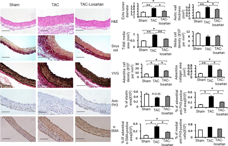
Left side: Representative cross sections of ascending aortas from sham-operated, TAC mice for 2 weeks, and TAC mice treated with losartan for 2 weeks by H&E staining, Sirius red staining for collagens, Verhoeff-van Gieson (VVG) staining for elastin, anti-phospho-histone H3 (PH3) staining for proliferative cells and anti-smooth muscle α-actin (α–SMA) staining for smooth muscle cells separately. Right side: Bar graph show mean ascending aortic lumen diameters of sham-operated mice (white bars), TAC mice (black bars) and TAC mice treated with losartan for 2 weeks (gray bars) (n = 5 per group). Bar graphs show quantification of aortic wall thickness, total media area, medial cell density, adventitial cell density, adventitial collagen area, percent elastin area, percent adventitial anti-PH3 positive area, percent medial anti-PH3 positive area and percent adventitial anti-α-SMA positive area from sham-operated mice (white bars), TAC mice (black bars) and TAC mice treated with losartan for 2 weeks (gray bars). n = 5 per group. *, P<0.05. **, P<0.01.