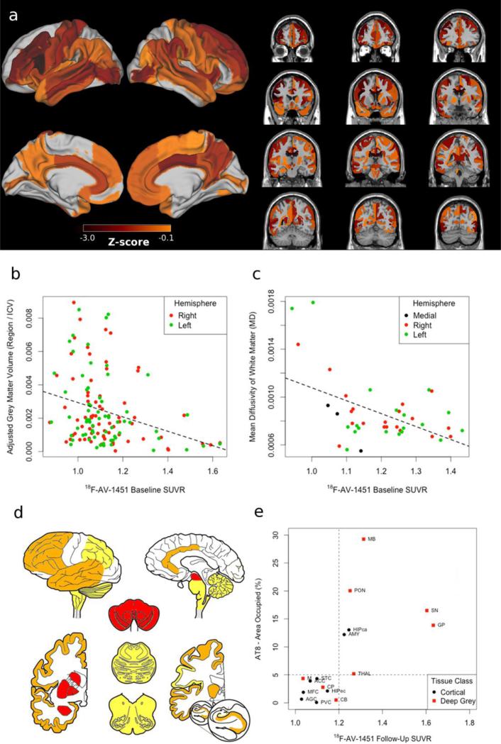
Figure 2. MRI/DTI neuroimaging, Histopathology and 18F-AV-1451.
a) Z-scores of grey matter MRI and white matter DTI relative to 100 demographically-comparable healthy controls; correlations demonstrating increased baseline (15 months pre-death) 18F-AV-1451 retention associated with reduced b) grey matter volume and c) white matter integrity; d) heatmap reflecting ordinal ratings of PHF-1 tau burden rated as severe (3+; red) in midbrain (MB), pons (PON), substantia nigra (SN), globus pallidus (GP), putamen (CP), and amygdala (AMY); moderate (2+; orange) in thalamus (THAL), medulla (M), and CA1/subiculum (HIPca) along with anterior cingulate (ACC), middle frontal (MFC), entorhinal (HIPec), angular gyrus (AGC), superior temporal (STC) cortices; mild scattered tangles (1+; yellow) in cerebellum (CB); and no pathology in primary visual cortex (PVC); e) Correlation between AT-8 percent area occupied and follow-up (5 months pre-death) 18F-AV-1451 retention.

