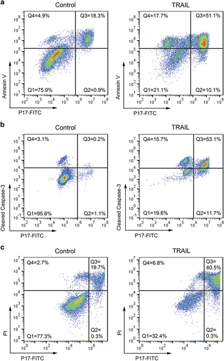
Figure 6.
Comparison of P17 with conventional apoptosis detection agents in apoptotic cells. TRAIL-treated and control Jurkat cells were incubated with FITC-P17 for 30 min, and then co-stained with Annexin V (Alexa Fluor 647-conjugated; a), cleaved caspase-3 antibody (Alexa Fluor 647-conjugated; b) or PI (c), before analysis on the flow cytometer. All the samples were plotted as P17 on the x axis versus other apoptosis-imaging agents' fluorescence on the y axis. The quadrants Q in (a) and (b) were defined as Q1=live (P17-negative/Annexin V- or CC3-negative), Q2=necrosis (P17-positive/Annexin V- or CC3-negative), Q3=late apoptosis (P17-negative/Annexin V- or CC3-positive) and Q4=early apoptosis (P17-positive/Annexin V- or CC3-negative). The quadrants in (c) were defined as Q1=live (P17-negative/PI-negative) and Q3=late apoptosis (P17-positive/PI-positive).

