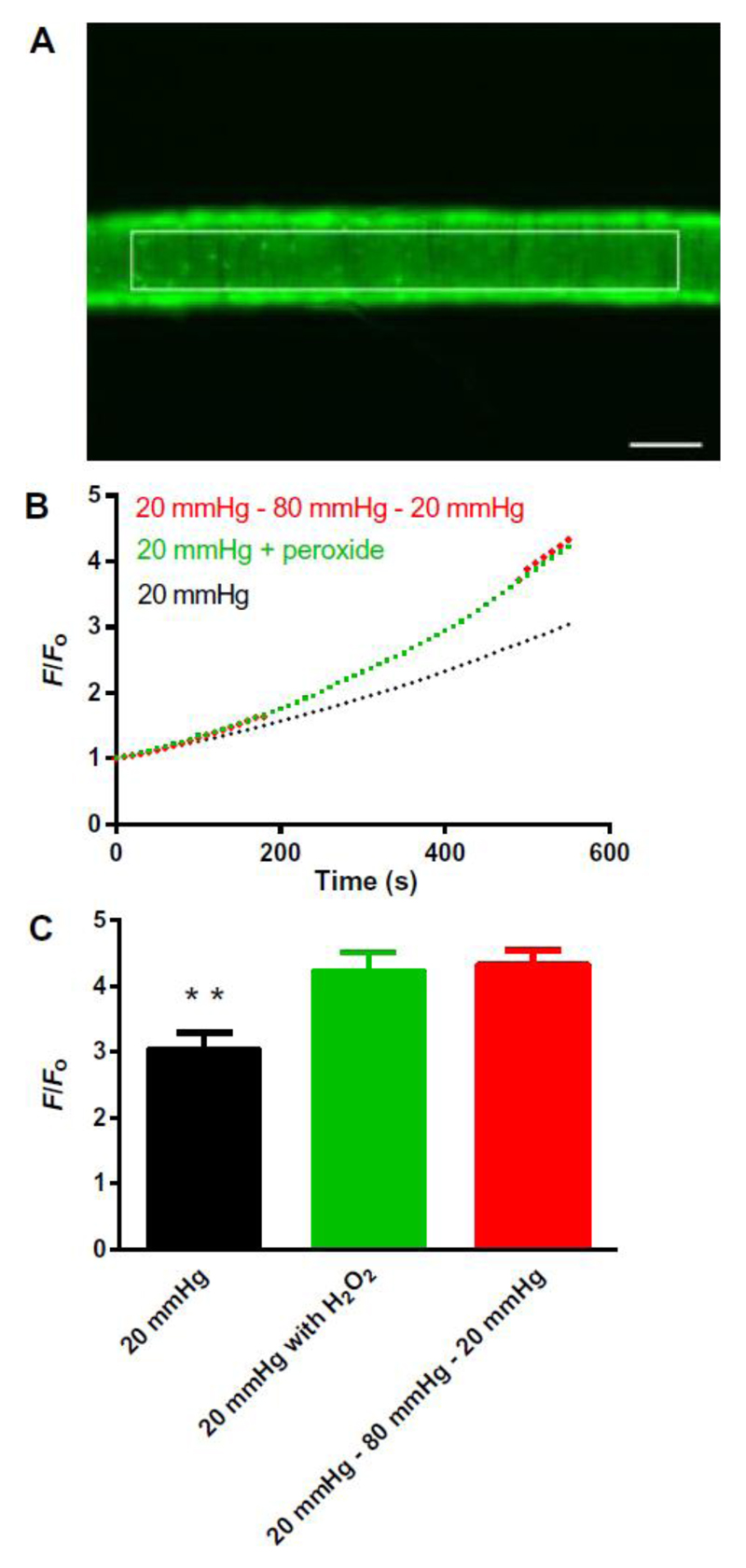
Figure 2. Intraluminal pressure increases oxidant amounts in mesenteric arteries.
A: Representative example of a third-order mesenteric artery loaded with CM-DCF and pressurized at 20 mmHg showing the region of interest (ROI). White scale bar: 100 μm. B: Averaged traces (SEM bars omitted) showing the rate of the increase in fluorescence in CM-DCF–loaded WT arteries continuously pressurized to 20 mmHg (black line; n = 4 arteries from 4 mice); continuously pressurized to 20 mmHg with exogenous application of the antioxidant H2O2 (30 μM), added at 240 seconds (red line; n = 4 arteries from 4 mice); and pressurized initially to 20 mmHg and then stepped to 80 mmHg at 210 seconds, and back to 20 mmHg at 480 seconds (green line: n=4 arteries from 4 mice). Dotted green line reflects artifactual changes in fluorescence caused by movement of the artery out of the focal plane. Representative traces for each protocol are shown in Supplementary Figure 3. C: End point (at 10 minutes) comparisons of increases in F/F0 for control arteries continuously pressurized to 20 mmHg (F/Fo = 3.0 ± 0.3; n = 4 arteries from 4 mice), continuously pressurized to 20 mmHg with exogenous addition of H2O2 (F/Fo = 4.2 ± 0.3; n = 4 arteries from 4 mice), and subjected to a pressure-step protocol from 20 to 80 to 20 mmHg (F/Fo = 4.3 ± 0.2; n = 4 arteries from 4 mice), presented as means ± SEM. *P = 0.01, compared with control at 20 mmHg.

