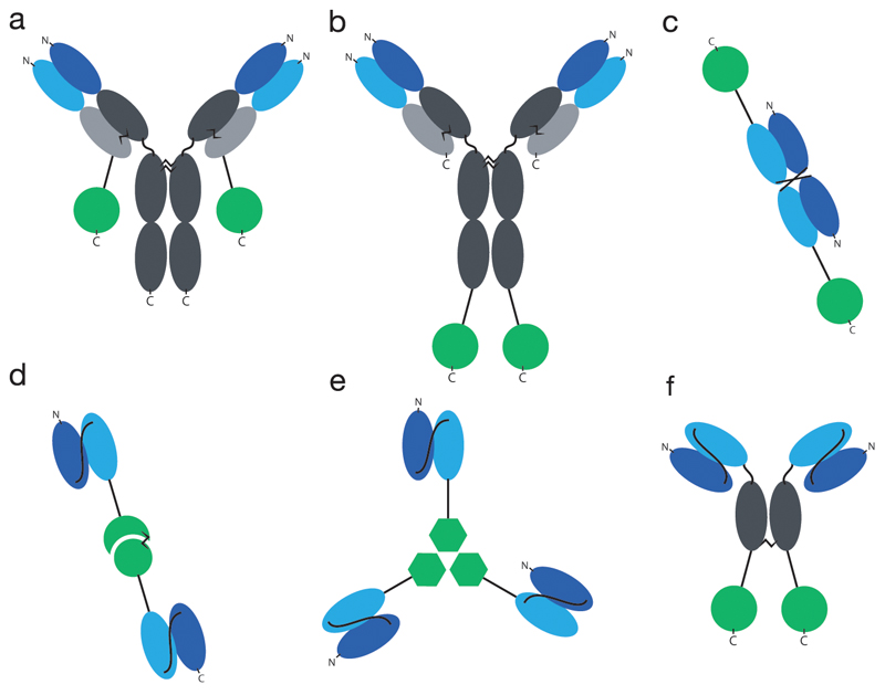
Fig. 1. Overview of common formats for immunocytokines.
a) IgG format, cytokine fused to the light chain, b) IgG format, cytokine fused to the heavy chain, c) Diabody, d) bivalent scFv format, here in fusion with heterodimeric cytokine IL12, e) trivalent scFv format, here in fusion with trimeric cytokine TNF, f) SIP format. Constant regions indicated in grey, VH indicated in dark blue, VL indicated in light blue, cytokines indicated in green (circle: monomeric cytokine like e.g. IL2, circle connected to half-circle: heterodimeric cytokine like e.g. IL12, hexagon: homotrimeric cytokine like e.g. TNF).

