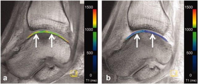
Figure 5.
Delayed gadolinium-enhanced magnetic resonance imaging of the talus after matrix-associated autologous chondrocyte implantation (MACI) at 3 T. White arrows indicate repair tissue borders. Image (a) displays precontrast T1-maps. Image (b) displays postcontrast T1-maps.46

