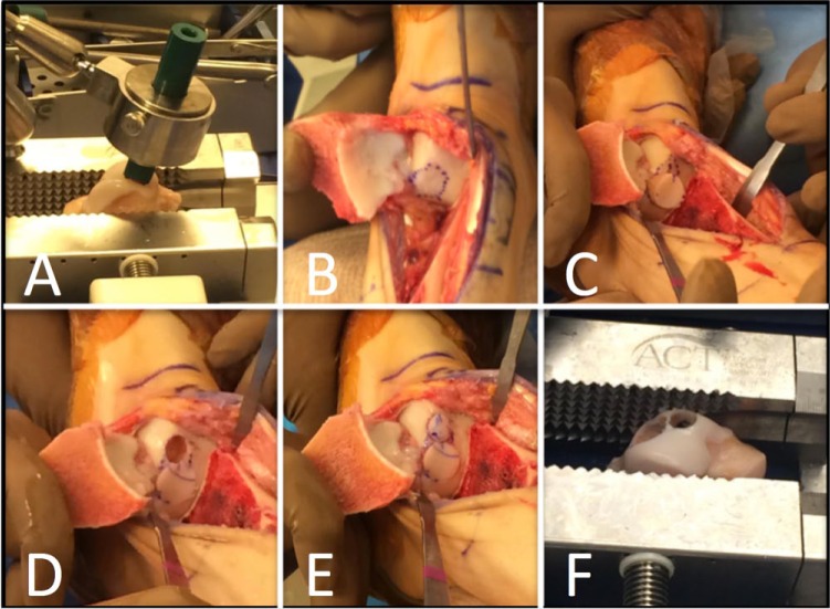
Figure 3.
Intraoperative photographs of a failed debridement and microfracture for a large talar dome lesion. (A) A size-matched cadaveric talus is secured within a transplant vice. (B) After medial malleolar osteotomy the lesion is circumscribed and sized for allograft. (C) The larger allograft plug is placed first and the remaining lesion is circumscribed for second plug using the “snowman” technique. (D) The talus is prepared for the second plug. (E) Both plugs have been press-fit into the defect. (F) The cadaveric talus after donation of both osteochondral plugs.

