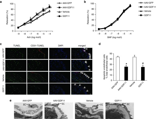
Figure 2.
AAV-GDF11 and GDF11 improved endothelial function and decreased endothelial cells apoptosis in apoE−/− mice. Four thoracic aorta rings from each apoE−/− mice (n = 3 mice from each group) were equilibrated for 1 hour at a preload tension of 0.5 g in Krebs buffer and then precontracted with norepinephrine (NE, 10–6 mmol/l). Once a steady state was achieved, vasodilation responses were evaluated by cumulative concentration-response curves to (a) acetylcholine (ACh, 10–9–10–4 mmol/l) and (b) sodium nitroprusside (SNP, 10–9–10–4 mmol/l). (c) Five continuous sections from each apoE−/− mice (n = 4 mice from each group) were costained with terminal deoxynucleotidyl transferase-mediated dUTP-biotin nick end labeling (TUNEL) (apoptotic cells, green), anti-CD31 (endothelial cells, red) and 4′,6-diamidino-2-phenylindole (DAPI) (nuclei; blue). (d) The percentage of apoptotic endothelial cells per total endothelial cells. (e) Electron microscopy was performed on thoracic aortas using ultrathin sections and examined with a Nikon EclipseE800 light microscope. Arrow shows endothelial cell (EC), IEL: internal elastic lamin. Scale bar, 20 μm. Data were shown as mean ± SD. Differences between 2 groups were tested with Student's t-test. *P < 0.05 versus AAV-GFP; #P < 0.05 versus vehicle group. AAV, adeno-associated viruses; AAV-GDF11, AAV-mediated GDF11 gene transfer; apoE−/−, apolipoprotein E null; GDF11, growth differentiation factor 11; GFP, green fluorescent protein; SD, standard deviation.

