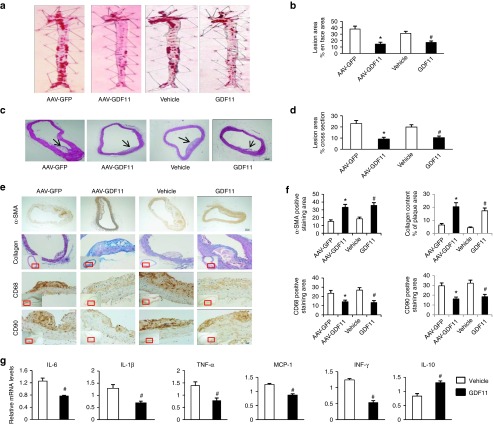
Figure 3.
AAV-GDF11 and GDF11 attenuated atherosclerotic lesion formation, ameliorated plaque components and inflammatory cytokines in aortas of apoE−/− mice. (a) Oil red O staining of the whole aorta (n = 4 mice in each group) was performed to analysis the en face atherosclerotic lesions area. (b) Quantitative analysis of a. (c) Hematoxylin and eosin staining (n = 4 mice in each group) was performed to quantify luminal cross-sectional area involved by atherosclerotic plaque. (d) Quantitative analysis of c. (e) Immunohistochemical staining of α-SMA, anti-CD68, and anti-CD90 were performed to analysis the VSMCs, macrophages and T lymphocytes expression levels. Scale bar, 20 μm. Masson's trichrome staining was performed to quantification of collagen content. Scale bar, 100 μm. (f) Quantitative analysis of e. (g) mRNA expression levels of IL-6, IL-1β, TNF-α, MCP-1, IFN-γ, and IL-10 measured by quantitative real-time polymerase chain reaction (PCR) in the abdominal aorta (n = 6 mice in each group) and expressed relative to β-actin. Data were shown as mean ± SD. Differences between two groups were tested with Student's t-test. *P < 0.05 versus AAV-GFP; #P < 0.05 versus vehicle group. AAV, adeno-associated viruses; AAV-GDF11, AAV-mediated GDF11 gene transfer; apoE−/−, apolipoprotein E null; GDF11, growth differentiation factor 11; GFP, green fluorescent protein; TNF-α, tumor necrosis factor-α; IL, interleukin; IFN-γ, interferon-γ; VSMCs, vascular smooth muscle cells; SD, standard deviation.

