Figure 5.
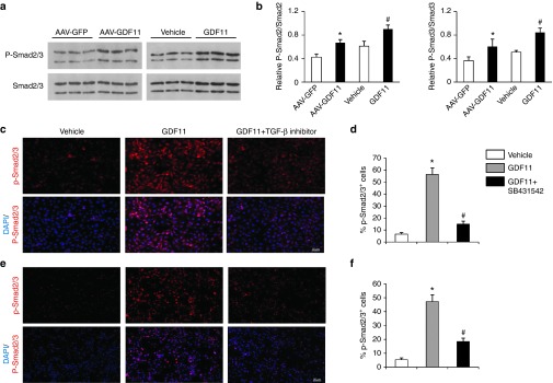
GDF11 activated TGF-β/Smad pathways in vivo and in vitro. (a) Expression of Smad2/3 and phosphorylated Smad2/3 in the aortas of apoE−/− mice were determined by western blot 12 weeks post GDF11 or others treatments. (b) Quantitative analysis of a. Data were shown as mean ± SD. Differences between two groups were tested with Student's t-test. n = 3 mice in each group. *P < 0.05 versus AAV-GFP; #P < 0.05 versus vehicle group. (c and e) Representative images of the percentage of phosphorylated-Smad2/3+cells in c MAECs and e RAW 264.7 macrophages treated with either GDF11 (50 ng/ml) for 30 minutes or pretreated with SB431542 (10 μmol/l) for 30 minutes, and then treated with GDF11(50 ng/ml) for 30 minutes. (d and f) Quantitative analysis of c and e. Data were shown as mean ± SD. Analysis of variance (ANOVA) followed by LSD t-test was used to compare the differences among different groups. Each experiment repeated five times. *P < 0.05 versus vehicle; #P < 0.05 versus GDF11 group. AAV, adeno-associated viruses; AAV-GFP, AAV-green fluorescent protein; GDF11, growth differentiation factor 11; MAECs, mice aortic endothelial cells; SD, standard deviation; TGF-β, transforming growth factor; LSD, least significant difference.

