Figure 5.
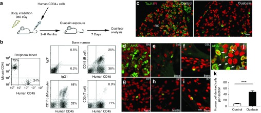
Enhanced tissue engraftment of human CD34+ cord blood cells in ouabain-treated ANs in a humanized transplantation model. (a) Experimental approach using humanized mice and AN injury. (b) Multi-lineage hematopoietic reconsititution from CD34+ cord blood cells. The data shows flow cytometric analysis of nucleated peripheral blood and bone marrow cells obtained from a representative NSG mouse six months after injection with 2.0 × 10⁵ CD34+ human cord blood cells. Reconstitution by human CD45+ cells in mouse peripheral blood was 24%. Reconstitution in bone marrow by human CD19+ (B cells), CD11b+ (monocytes) and CD3+ (T cells) was 25, 18, and 0.9%, respectively. (c) Ouabain destroys almost all SGNs (Tuj1+) in the cochlea of a representative transplanted NSG mouse. (d) Abundant human cells identified by immunostaining with anti-human antigen (AHA) are present in the BM. (e–i) Human cells derived from cord blood HSCs (AHA+ cells) are present in several locations in the cochlea of a mouse 5 months after transplantation. The images were obtained 7 days after exposure of the cochlea to ouabain and include the spiral ligament (SpL; e), the osseous spiral lamina (OSL; f) and Rosenthal's canal (RC; g–i) from the apical, middle, and basal turns. (j) Human cells (human lung cancer cell line A549) were cocultured with mouse cells (mouse auditory neuroblast cell line N33). Human cell nuclei (white arrows) are stained green with AHA, whereas nuclei of mouse cells remain unlabeled (white arrowheads). (k) The tissue engraftment of human HSC-derived cells increased significantly in the AN of ears exposed to ouabain. Data was obtained from the left (control) and right (ouabain exposed) ears of NSG mice (***P < 0.001; data represent mean ± SEM; n = 5 mice per group; P = 0.00005; t = 18.14; unpaired t-test). Scale bars = 25 µm in c; 10 µm in i (applicable to d–h); 7 µm in j.

