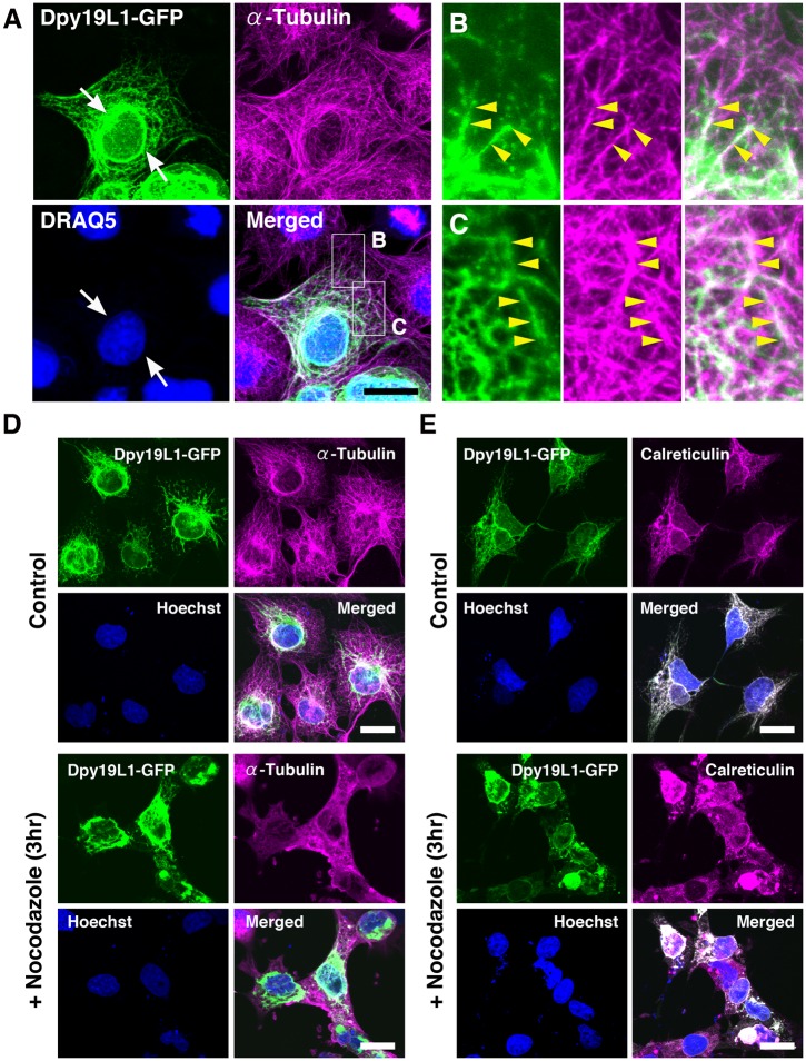
Fig 2. Distribution patterns of Dpy19L1 along microtubules in COS-7 cells.
(A-C) Double staining of Dpy19L1-GFP (green) and endogenous α-Tubulin (magenta) in COS-7 cells transfected with Dpy19L1-GFP. Nucleus was labeled by DRAQ5 (blue). Lower right panel shows the merged image. Boxed areas are magnified in B and C. Dpy19L1 is highly localized in a perinuclear region (arrows). The meshwork-like pattern of Dpy19L1 along the microtubule network is observed (yellow arrowheads). (D,E) COS-7 cells transfected with a pDpy19L1-GFP plasmid were treated by nocodazole, an inhibitor of microtubule assembly, for 3 h before fixation. (D) Immunostaining of GFP and α-Tubulin. (E) Immunostaining of GFP and Calreticulin. The cytoplasmic reticular staining of Dpy19L1 is severely disrupted by application of nocodazole. Nucleus was labeled by Hoechst 33342 (blue). Results shown here were obtained from at least three independent culture experiments. Scale bars: 20 μm.

