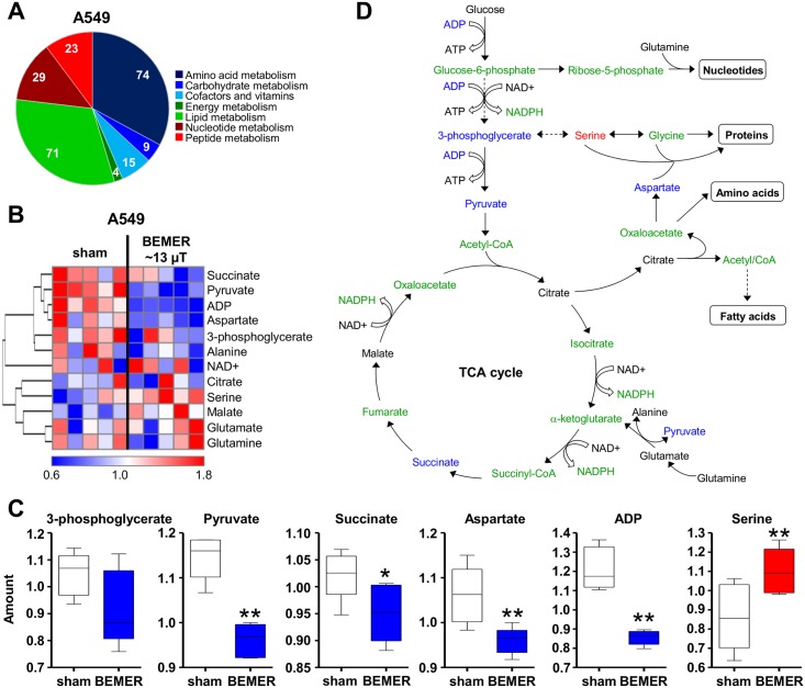
Fig 2. The specific BEMER EMF pattern impacts on cancer cell metabolism.
(A) Pie chart showing the number of detected metabolites categorized by pathways (Σ 225). (B) Heatmap comparing levels of metabolites in BEMER signal treated (~13 μT, 8 min) and BEMER sham-treated (sham) A549 cells. Red and blue indicate up- and downregulation, respectively. Cells were cultured in 3D lrECM for 24 h prior to BEMER treatment. (C) Amount of indicated metabolites in A549 cells without (sham) and with BEMER EMF exposure. (D) Scheme of glycolysis and TCA cycle. Metabolites in blue were downregulated, in red upregulated and in black unaffected upon BEMER therapy compared with sham-treated controls. Metabolites depicted in green were not measured in the metabolome analysis. All results represent mean ± SD. Student's t-test. n = 5. * P < 0.05; ** P < 0.01.

