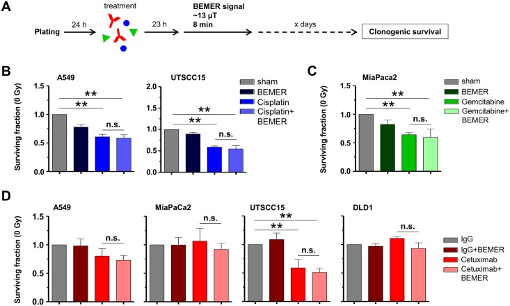
Fig 6. Sensitivity to chemotherapy and Cetuximab is not influenced by BEMER therapy.
(A) Flow chart of colony formation assay. Cells were plated in 3D lrECM, treated with respective agents followed by BEMER therapy 23 h later. (B) Basal surviving fraction after Cisplatin (0.1 μM; DMEM as control) treatment and BEMER therapy (~13 μT, 8 min). (C) Basal surviving fraction after Gemcitabine (10 nM; DMEM as control) treatment and BEMER therapy (~13 μT, 8 min). BEMER sham-treated (sham) cells served as control. (D) Basal surviving fraction after Cetuximab (5 μg/ml; IgG as control) treatment and BEMER therapy (~13 μT, 8 min). IgG-treated cells served as control. All results represent mean ± SD. Student's t-test. n = 3. * P < 0.05; ** P < 0.01. n.s., not significant.

