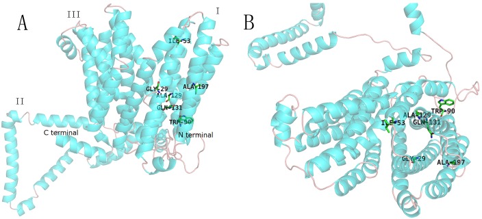
Fig 3. The modeled 3D structures of AmpG from PAO1.
Twelve TM helices and one internal membrane region are illustrated. The first and third parts consist of six transmembrane α helices. The region between the first and third parts (named the second part) is a sequence of 167 AA in length. The mutated amino acids at positions 29, 53, 90, 129, 131 and 197 are illustrated in the third part (A). The picture on the right illustrates the protein as viewed from outside of the membrane (B).

