
Fig. 4.
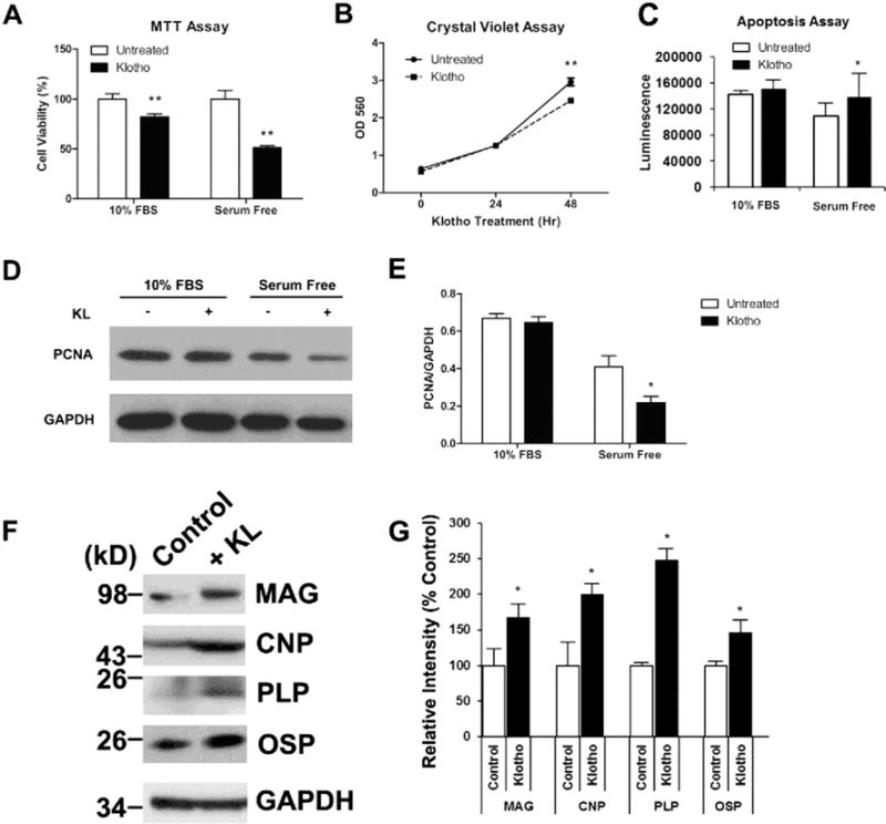
Klotho inhibits MO3.13 cell proliferation, increases apoptosis, and enhances differentiation. a MO3.13 cells were treated with Klotho for 48 h in either DMEM containing 10 % FBS or serum-free DMEM and assayed for cell proliferation using the MTT assay. Cells treated with Klotho are 15 and 48 % less confluent when grown in DMEM containing 10 % FBS or serum-free DMEM, respectively. b Cells treated with Klotho for 48 h in DMEM containing 10 % FBS show a significant decrease in cell number as measured by the Crystal Violet assay. *p< 0.001 indicate statistical significance by t test. Error bars indicate standard deviation. Results are from three to five independent experiments. c MO3.13 cells were treated with the same conditions as in a for 48 h and assayed using the cell apoptosis assay. Cells treated with Klotho have a significant increase in apoptosis when grown in serum-free DMEM. d Western blot analysis of the same experiments in a using an antibody against the cell proliferation marker PCNA with GAPDH as loading control. e Statistical analysis of densitometric results from d. *p<0.05 indicate statistical significance by t test. Results from a–d represent the average of three to five independent experiments. f MO3.13 cells were treated with Klotho for 7 days in DMEM OPC culture medium as described in “Materials and Methods” section. The cell lysates were collected for SDS-PAGE using antibodies against mature oligodendrocyte markers indicated, with GAPDH as control. g Statistical analysis of densitometric results from f. *p<0.05 indicate statistical significance by t test. Results are from three independent experiments