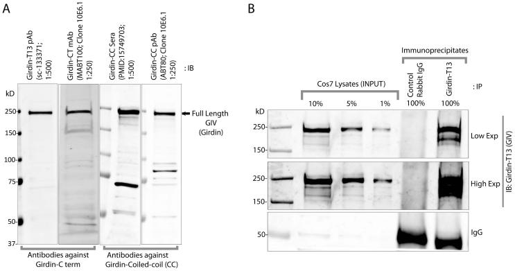
Figure 2. Detection of GIV/Girdin by immunoblotting and immunoprecipitation.
A: Equal aliquots of lysates (~ 60 μg) of COS-7 cells were analyzed for GIV expression by immunoblotting (IB) with a variety of commercially available monoclonal and polyclonal antibodies raised against either the coiled-coil (N-terminal) or C-terminal (CT) domains of GIV at the indicated dilutions. Full-length GIV is detected at ~250 kD by all antibodies. In addition, some coiled-coil antibodies detect a few shorter products in a variety of cell lines, some of which are isoforms of GIV. B. Equal aliquots of lysates (~ 2.0 mg) of COS-7 were incubated with ~1.5 μg control or anti-Girdin T13 (SCBT) antibody and protein A beads. Immune complexes were eluted and analyzed for GIV by IB.

