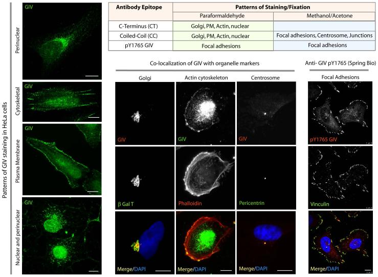
Figure 4. Subcellular distribution of GIV, as determined by whole cell immunofluorescence.
HeLa cells grown on cover slips were fixed and co-stained for GIV with a variety of other markers (as indicated) and analyzed by confocal immunofluorescence. Table 2 lists the commercially available anti-GIV antibodies, and the patterns of staining seen using each antibody.

