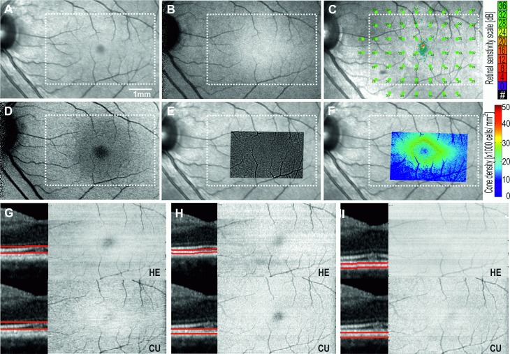
Fig 3.
Multimodal imaging of the left eye of a healthy subject showing normal 15° × 10° Near-infrared reflectance NIR (A), infrared autofluorescence IRAF (B), microperimetry (C), blue-light autofluorescence BAF (D), adaptive optics flood illumination ophthalmoscopy AO-FIO cone montage and density maps overlaid on NIR (E,F), and en face OCT maps of the ellipsoid zone EZ (G), interdigitation zone IZ (H), and retinal pigment epithelium RPE (I). HE, Heidelberg Eye Explorer software (Heidelberg Engineering); CU, Custom-built software using graph-search theory algorithm. White dotted rectangle corresponds to region for which en face maps are generated.

