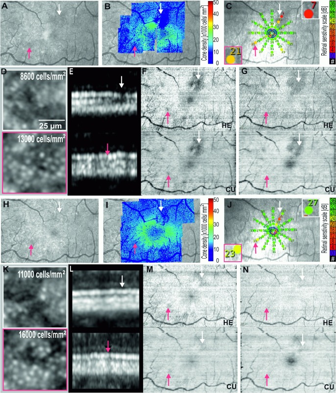
Fig 5.
Multimodal imaging showing the left eye of Case 2: acute macular neuroretinopathy at presentation (A-G), and 6 months (H-N) of follow-up. Near-infrared reflectance NIR (A,H), adaptive optics flood illumination ophthalmoscopy AO-FIO density map overlaid on NIR (B,I,) and magnified cone images (D,K), microperimetry (C,J), OCT B-scans in the region of relative scotoma (E,L) and en face OCT images of ellipsoid (F,M), and interdigitation zones (G,N) showed partial recovery. Both commercial and custom software were able to demonstrate the topography of ellipsoid and interdigitation zone injury.

