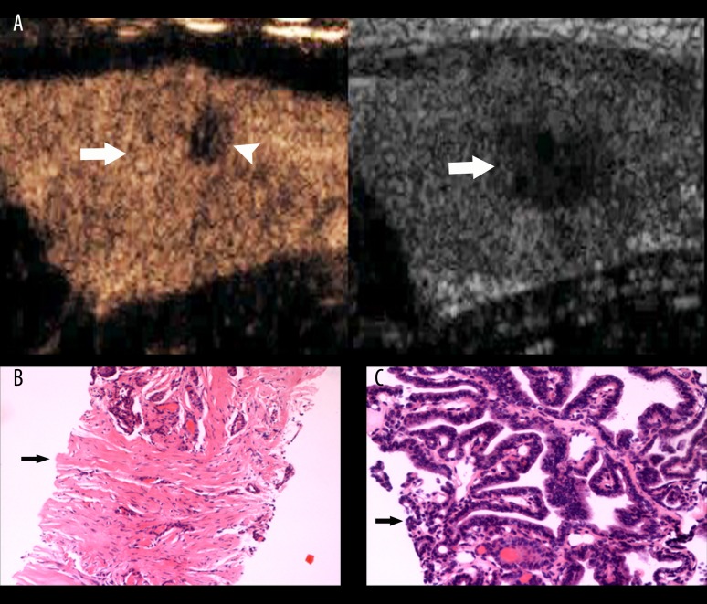
Figure 2.
Thyroid papillary carcinoma. (A) A hypoechoic lesion (arrow) with unclear boundary and irregular shape was found in the left lobe of thyroid by conventional US (right image). Iso-enhancement with focal low-enhancement (arrow head) was seen by CEUS. No ring was seen in its peripheral area (left image, arrow). (B) Many fibers, a few blood vessels (arrow), and a few cancerous cells were apparent in a biopsy specimen from an area of low-enhancement. (C) A biopsy specimen from an area of iso-enhancement showed many cancerous cells (arrow).

