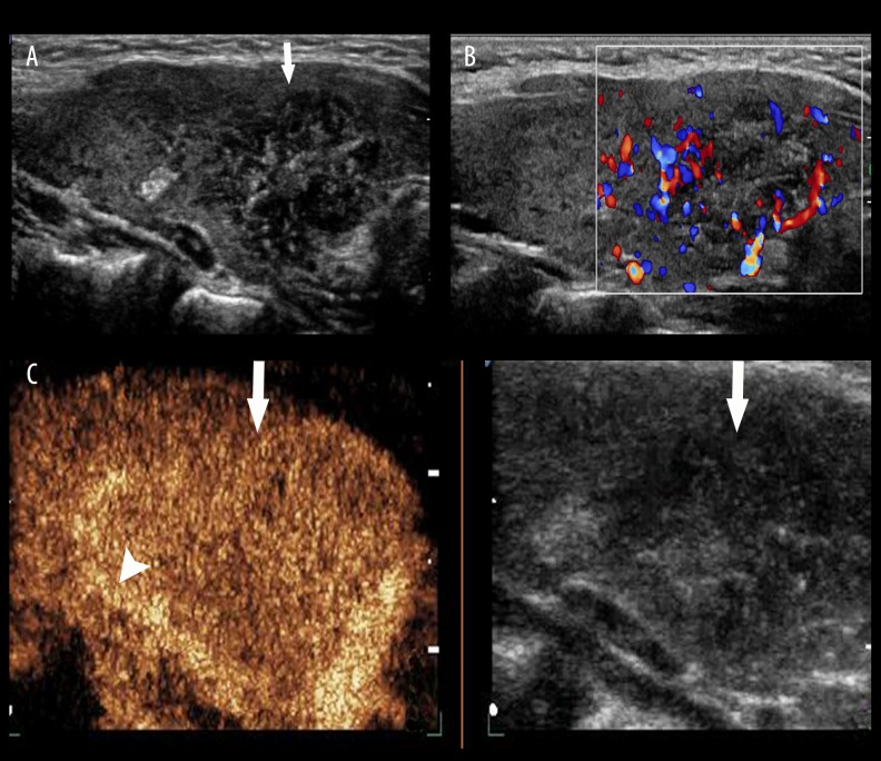
Figure 4.
Thyroid papillary carcinoma. (A) A hypoechoic lesion (arrow) with ill-defined boundaries and a lot of microcalcifications was found in the left lobe of the thyroid gland by conventional US. (B) Rich blood signals were detected in lesion by CDFI. (C) Heterogeneous enhancement was seen in the lesion, and an irregular high-enhanced ring (arrow head) was shown in the periphery by CEUS (arrow).

