Figure 5.
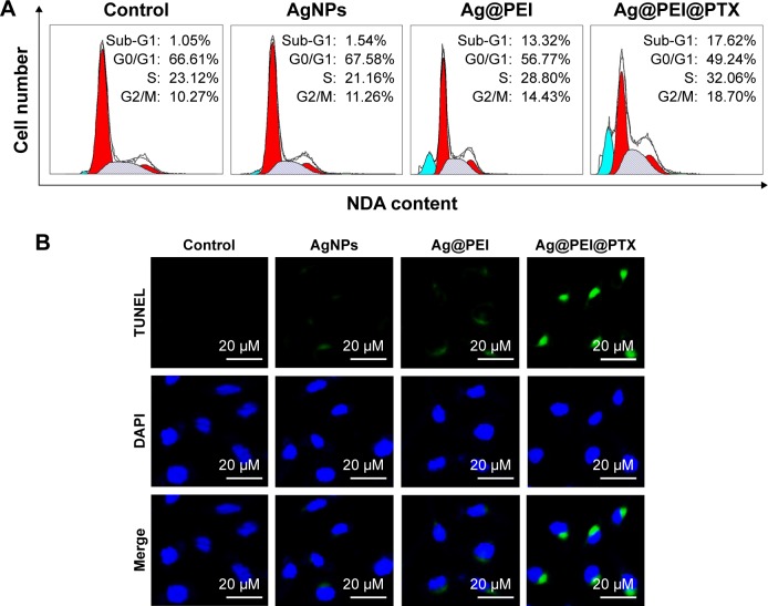
Ag@PEI@PTX-induced apoptosis in HepG2 cells.
Notes: (A) The cell-cycle distribution with different treatments was analyzed by quantifying DNA content using flow cytometric analysis. (B) Representative photomicrographs of DNA fragmentation and nuclear condensation as detected by TUNEL–DAPI co-staining assay.
Abbreviations: AgNPs, silver nanoparticles; DAPI, 4′6-diamidino-2-phenylindole; TUNEL, terminal transferase dUTP nick end labeling.

