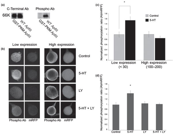
Fig. 9.
PKC Apl III phosphorylated at the PDK site is increased after 5HT treatment. (a) 10 ng of purified GST-catalytic domain and 50 ng of SF9 cell extract from cells infected with a baculovirus expressing PKM Apl III were separated on SDS–PAGE acrylamide gels, transferred to nitrocellulose, and immunostained with the antibody to the carboxyterminal (left) or the phospho-specific antibody to the PDK site (right). (b) Sensory cells over-expressing PKC Apl III were treated with either a control solution, 5HT (20 μM for 10 min), LY 294002 (10 μM for 10 min), or both LY and 5HT: LY (10 μM) was applied for 15 min prior to the 5HT (10 min) treatment. Cells were immediately fixed and immunostained with the phospho-specific antibody and imaged for mRFP. Examples are shown either at low levels of expression or high levels of expression. (c) Quantification of normalized phosphorylation ratio for low and high expressing neurons: low expression data from three experiments, C, n = 14, 5HT, n = 22, and high expression from two experiments, C, n = 7, 5HT, n = 8. *, p < 0.05 Students two-tailed paired t-test using non-normalized data. (d) Quantification of normalized phosphorylation after experimental treatments (C, five experiments, n = 18; 5HT, four experiments, n = 44; LY three experiments, n = 12; 5HT + LY three experiments, n = 26). ANOVA F(96,3) = 8.9, p < 0.01; Tukey’s post-hoc test showed that 5HT was different from all other groups, *, p < 0.05 and no other differences were seen.

