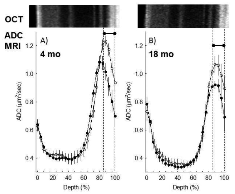
Figure 3. Light-evoked increase in SRS as measured by diffusion weighted MRI does not change with age.

Summary of central retinal ADC as a function of retinal depth during dark (closed symbols) and at 20 minutes of ∼500 lux light (open symbols) in A) 4 mo (n = 7) and B) 18 mo UM-HET3 (n = 5) mice. Data are presented using the conventions in Figure 2. *Retinal depth range with significant difference (P < 0.05).
