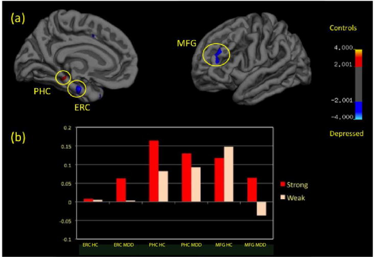
Figure 3.

Compared to Healthy Controls (HC), Depressed Patients (MDD) display reduced parahippocampal cortex (PHC) but increased entorhinal cortex (ERC) and medial frontal gyrus (MFG) differential activity during contextual processing (p<0.01). (a) Displays the distribution of this differential activity and (b) displays the levels of activation (percent signal change) in these regions during the Strong (red) and Weak (white) contexts.
