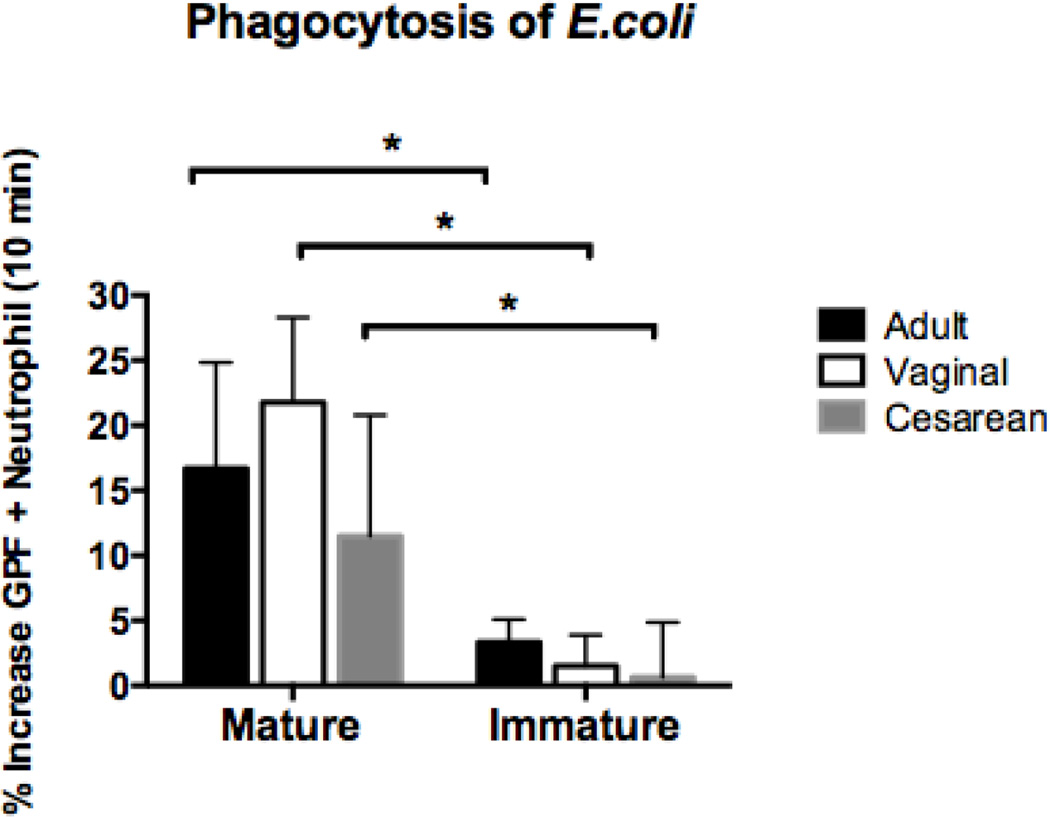
Figure 2. Phagocytosis of E.coli based on neutrophil maturity in adults, neonates born vaginally, and neonates delivered by primary cesarean section.
GFP-labeled E.coli (RS218; 1 × 10⁸ organisms/ml) were added to whole blood in sodium heparin tubes from vaginal (n=4), cesarean (n=5), and adult (n=7) samples. Duplicate samples were incubated at 37°C for 10 minutes. Immediately after the incubation period, the samples were transferred to ice and labeled with CD11b and CD16. Flow cytometry was used to measure mean fluorescence intensity of the neutrophils following ingestion of GFP- labeled E.coli. Mature cells were defined by a population of cells high in CD11b (APC) and CD16 (PE), and consisted of segmented and banded neutrophils. Immature granulocytes had lower levels of CD11b (APC) staining and varied levels of CD16 (PE). Metamyelocytes, promyelocytes, and myelocytes comprised the immature granulocyte population. Mature neutrophils in neonates and adults phagocytosed E.coli equally well, while immature cells performed poorly. **p < 0.001.

