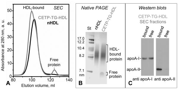
Figure 1.
Characterization of human plasma HDL enriched with TG. Incubation mixture containing CETP (~70 kDa), VLDL, and HDL (CETP-TG-HDL) was analyzed by (A) SEC using preparative-grade Superdex 16 XK column and (B) native PAGE (4-20% gradient, Denville Blue protein stain). HDL-bound and free protein fractions are indicated. The void volume in SEC (not shown) contains large VLDL particles (40-100 nm). (C) Immunoblots of the HDL-bound and free protein fractions, which were isolated by SEC from CETP-TG-HDL, show that the free protein released from HDL upon enrichment in TG contains apoA-I but no apoA-II.

