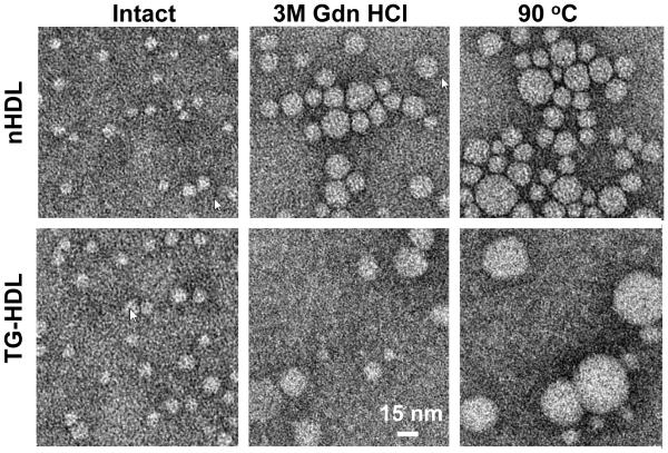
Figure 4.
Transmission electron micrographs of negatively stained nHDL and TG-HDL that were either intact (left), or have been perturbed by a denaturant (incubation with 3 M Gdn HCl at 37 °C for 3 h; middle), or by heating (to 90 °C at a rate of 90 °C /h, right). Representative images are shown on the same scale.

