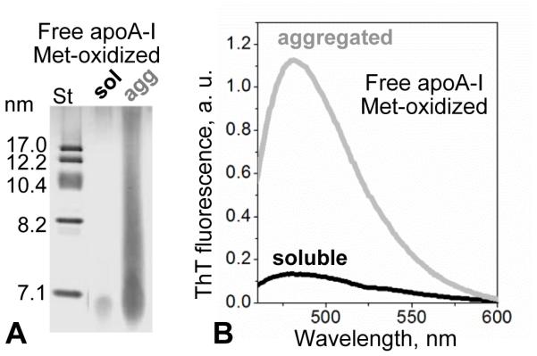
Figure 6.

Aggregation of Met-oxidized free apoA-I released upon mild oxidation of TG-HDL. Free protein, which was isolated from mildly oxidized TG-HDL by SEC as described in Figure 2 legend, was incubated at pH 6 as described in part 3.2 following published protocols [20]. (A) SDS PAGE showed massive protein aggregation upon incubation. (B) Thioflavin T fluorescence showed a large increase in emission in the presence of this aggregated protein, suggesting amyloid formation.
