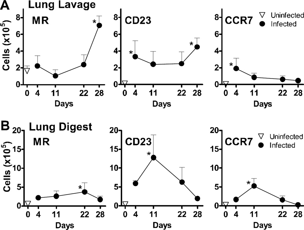
FIG 2.
Pneumocystis murina infection up-regulates the surface protein expression of alternative macrophage activation markers in vivo. Cells isolated from bronchoalveolar lavage (A) and lung digest (B) preparations from BALB/c mice isolated over time post-infection were enumerated and stained for cell surface expression of macrophage proteins and analyzed by flow cytometry. After gating out lymphocytes and dead cells by forward and side scatter characteristics, the number of cells within the CD11b+GR1− gate that exhibited up-regulation of MR, CCR7, and CD23 as compared to unstained controls were reported over time for each compartment. All graphs display mean ± SD for 3–4 mice per group per timepoint, and results are representative of 2 experimental replicates. Values were compared using 1-way ANOVA and Dunnett’s post-hoc analysis comparing means to that of an uninfected control group. Statistically significant differences between treatment groups indicated for P < 0.05 (*).

