
Figure 1. Overview of MAPK signaling and DUSP regulation.
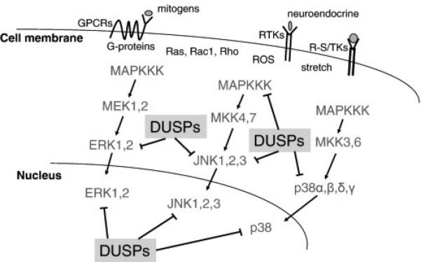
Simplified diagram of the MAPK signaling cascade showing each step in the cascade culminating in ERK1/2, JNK1/2/3 or p38α,β,δ,γ activation, which are inactivated by specific DUSP proteins in either the cytoplasm or nucleus. Other MAPK branches not shown include MEK5-ERK5 and ERK3/4, which are less well characterized for DUSPs counter-regulation.