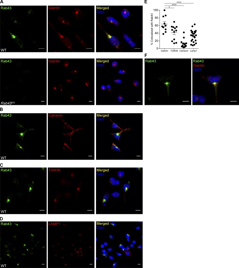
Figure 2.
RAB43 is abundant in Golgi and vesicles of Batf3-dependent DCs. (A) Day 10 FLT3L-cultured BM from WT (top) or Rab43Δ/Δ (bottom) 129 mice was treated with LPS for 4 h (to improve attachment), allowed to attach to coverslips, fixed, stained with 2E6 (anti-RAB43; green) and anti-giantin (red), and attached to slides using Prolong Gold antifade with DAPI (blue). (B–D) WT cells as described in A untreated with LPS were stained with 2E6 (green) and anti-calnexin (red; B), anti-TGN38 (red; C), or anti-LAMP1 (red; D). Coverslips were attached to slides using Prolong Gold antifade with DAPI (blue). (E) Percentage of organelle stain that is colocalized with RAB43 stain for the indicated organelles. Each dot represents a single cell with 10–23 cells analyzed per stain. Data were obtained using Imaris Coloc2. Statistics were analyzed using ANOVA. *, P < 0.05; ****, P < 0.0001. (F) WT cells prepared as described in A shown at increased zoom to highlight vesicular staining. All microscopy data are representative of at least two independent experiments. Bars, 5 µm.

