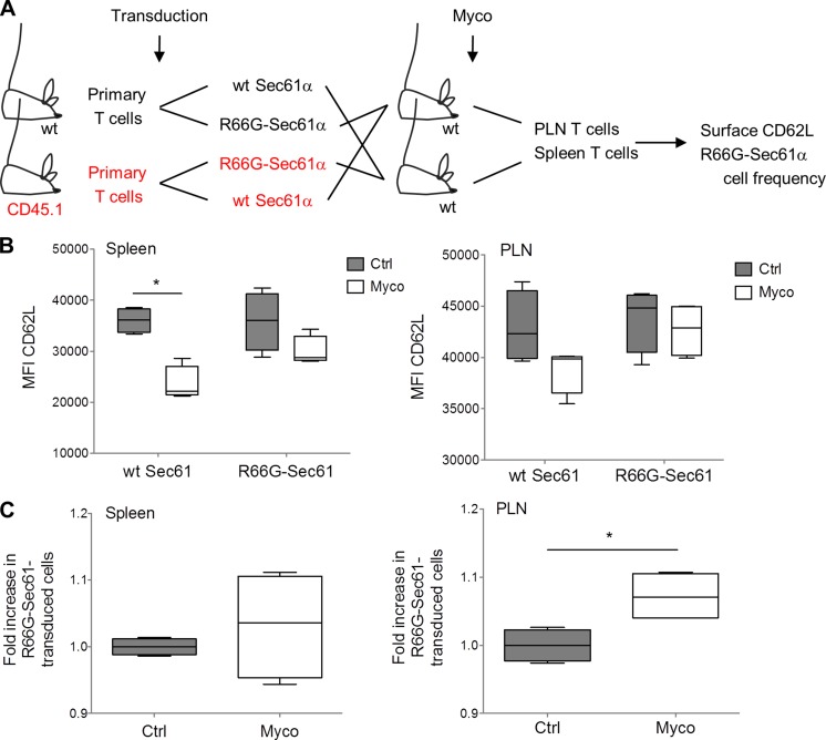
Figure 5.
Mycolactone suppresses Sec61 activity in T cells in vivo. (A) Primary T cells isolated from WT (C57BL/6J and CD45.2+) and congenic CD45.1 mice were transduced with WT Sec61α or R66G-Sec61α and mixed, as depicted. Each cell mix was injected intravenously into four recipient mice, two of which received concomitantly an intraperitoneal injection of mycolactone (Myco) and, the other two, vehicle as control. (B) CD62L surface expression on WT Sec61α and R66-Sec61α T cells (CD45.1+ or CD45.1−; Zsgreen+ gated) recovered from the spleen and PLN. Ctrl, vehicle control; MFI, mean fluorescence intensity. (C) Relative proportion of R66G-Sec61α cells, compared with WT Sec61α cells, in the spleen and PLN. (B and C) Data are mean fluorescence intensity (B) and mean cell numbers (C) in each experimental group, presented as box and whiskers (*, P ≤ 0.05, Mann-Whitney test, each box corresponding to four experimental values). They are representative of two independent experiments giving similar results.

