
Figure 8.
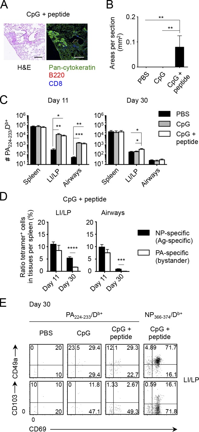
Local Ag plays a role in the formation of CD8+ TRM cells in the lung. (A–E) Mice infected i.p. with x31 were then injected i.n. with PBS, CpG ODN, or CpG ODN + NP366–374 peptide at day 8 as shown in Fig. 7 C. (A) Representative H&E-stained (left) and fluorescent (right) micrographs of peribronchiolar foci in mice treated i.n. with CpG ODN + NP366–374 peptide at day 30 PI. Pan-cytokeratin, green; B220, red; CD8, blue. Similar infiltrates were not observed in mice treated i.n. with PBS or CpG alone. Bars, 200 µm. (B) Mean areas of peribronchiolar foci in the lung were measured in mice shown in A at day 30 PI. (C) Numbers of PA-specific CD8+ T cells in each tissue at day 11 and 30 PI (day 3 and 22 after treatment). (D) Ratio of tetramer+ cells in tissues relative to those in the spleen ([# tetramer+ cells in a tissue/# tetramer+ cells in the spleen] × 100) of mice treated i.n. with CpG ODN + NP366–374 peptide. (E) Representative dot plots showing the expression of indicated molecules on PA- and NP-specific CD8+ T cells in the LI/LP of mice treated with either PBS, CpG ODN, or CpG ODN + NP366–374 peptide. Data are representative of two independent experiments (mean and SEM of five to six mice per group). *, P < 0.05; **, P < 0.01; ***, P < 0.001; ****, P < 0.0001 by one-way ANOVA with Tukey’s posthoc tests (B and C) and Student’s t test (D).