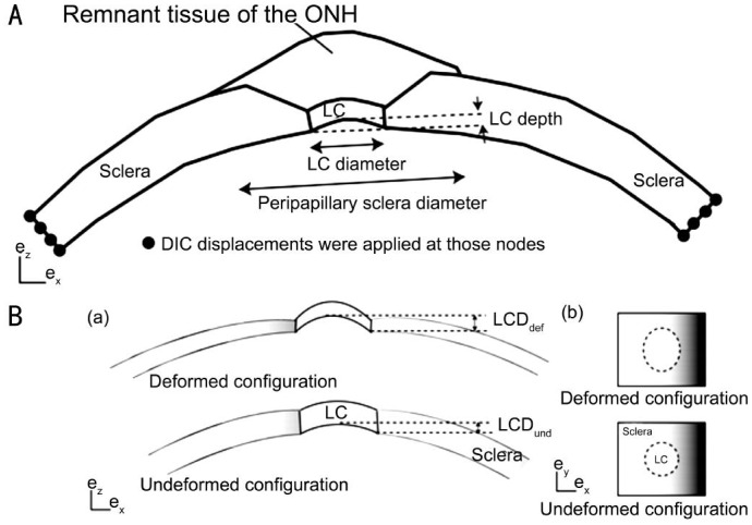
Figure 3. The finite element model of the posterior scleral and LC.
A: Schematic of the finite element model for the posterior scleral cup with the peripapillary sclera and the optic nerve head. The peripapillary sclera thins over a distance equal to the LC diameter; B: a) Schematic representation of the LC and the sclera in the (ex, ez) plane, showing the LCP; b) Schematic representation of the LC and sclera in the (ex, ey) plane.

