Figure 2.
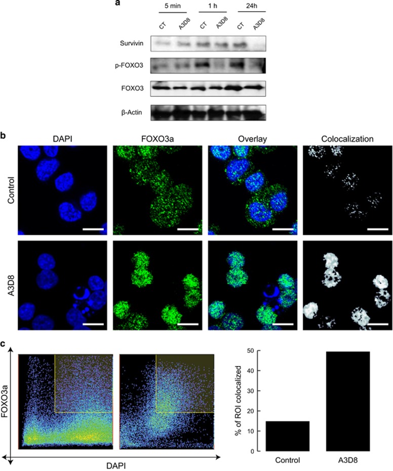
Significant anti-leukemic effects result following treatment of AML cells with anti-CD44-mAbs, which are coincident with reduction in markers of poor disease prognosis. (a) HL60 cells were cultured in presence of mIgG1 (CT) or of A3D8 (at 2.5 μg/mL) for the indicated times and subsequently cell lysates were subjected to western blot analysis using antibodies against survivin, p-FOXO3a (Ser253) and total FOXO3a protein. β-actin was used as a loading control. (b) A3D8 treatment leads to movement of FOXO3a out of the cytoplasm and into the nucleus. mIgG1-treated (CT) (upper panels) and A3D8-treated (lower panels). HL60 cells were fixed, permeabilized, and stained for total FOXO3a (green) and DAPI DNA nuclear stain (blue) prior to confocal fluorescence imaging. Colocalization mask of the green and blue channels is represented in white. One representative experiment of n=3 is shown. Scale bar, 10 μm. (c) HL-60 cells were treated with IgG1 (control) or A3D8 and immunofluorescence staining of FOXO3a was performed as well as nuclear staining using DAPI. Colocalization was analyzed from (b) with the nuclei as regions of interest (ROI). (Left) Dot plot of the distribution of pixel intensities within the ROI shows higher correlation between FOXO3a signal and DAPI signal compared with the control. (Right) The percentage of colocalized area was calculated in the ROI and showed higher colocalization in A3D8-treated cells (49.4%) compared with the control (14.8%). Z-stack analysis was also performed to show that the expression of FOXO3a protein predominated in the nucleus as illustrated in Supplementary Videos 1-8.

