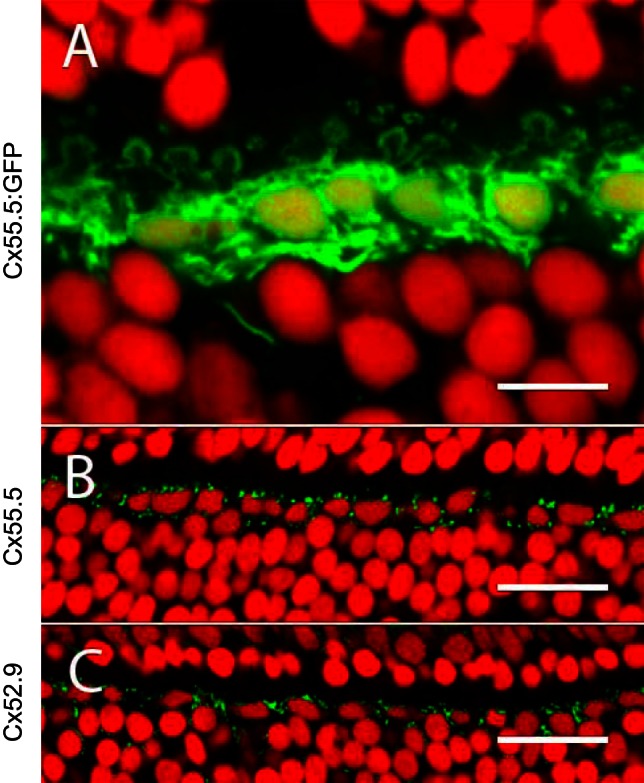
Fig. 3.

Immunohistochemical localization of zebrafish HC Cxs. A: section of a retina with GFP expression via the Cx55.5 promoter, showing a single layer of HC somata. The horseshoe-shaped structures are dendrites innervating cone synaptic terminals. Nuclei are stained with ethidium bromide (red). B and C: retinal sections stained with antibodies against Cx55.5 and Cx52.9 show punctuate labeling (green) around HCs. Nuclei are stained with ethidium bromide (red). Scale bars: 10 μm (A), 25 μm (B), 25 μm (C).
