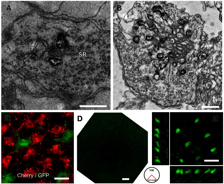
Fig. 5.
Specificity of GFP expression via the Cx52.7, Cx52.9, and Cx52.6 promoters. A: electron microscopic image of a rod synaptic terminal in a Cx52.7:GFP retina. HC dendrites flanking the synaptic ribbon (SR) are stained with an anti-GFP antibody. Scale bar: 500 nm. B: electron microscopic image of a cone synaptic terminal in a Cx52.7:GFP retina. HC dendrites flanking the multiple synaptic ribbons (SR) are stained with an anti-GFP antibody. C: confocal image of a double Cx52.7:GFP and Cx52.9:mCherry transgenic zebrafish shows that there is no overlap between the HC somata showing expression. The expression of mCherry led to some cluttering in the dendrites. D: overview of a Cx52.6:GFP retina. The ventral quadrant shows fluorescence only in HCs (H1 + H4); the dorsal 3 quadrants show fluorescence in H4 HCs and strong fluorescence in a specific type of bipolar cell. E: confocal image of the dorsal region of a Cx52.6:GFP retina, showing the morphology of bipolar cells with green fluorescence. Scale bars: 0.5 μm (A), 0.5 μm (B), 10 μm (C), 200 μm (D), 20 μm (E).

