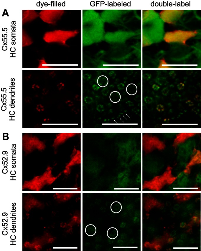
Fig. 8.

Morphological characteristics of biphasic HCs. A: filled HCs with a biphasic response profile (red) in a Cx55.5:GFP retina (green) showing an overlap of red and bright green HCs, indicating that the filled HCs are H2-type HCs. At the level of the dendrites, small rosettes show overlap of red and green fluorescence, while the largest rosettes (circles, indicating L-cone terminals) and dendrites inside rod terminals (arrows) were not filled. Scale bars: 10 μm. B: filled HCs with a biphasic response profile (red) in a Cx52.9:GFP retina (green) showing an alternating pattern of red and green fluorescent HCs, indicating that the filled HCs are not H1 type HCs. The largest rosettes (circles, indicating the L-cone terminals) show green but not red fluorescence, indicating innervation from H1-type HCs but not innervation from H2-type HCs. Scale bars: 10 μm.
