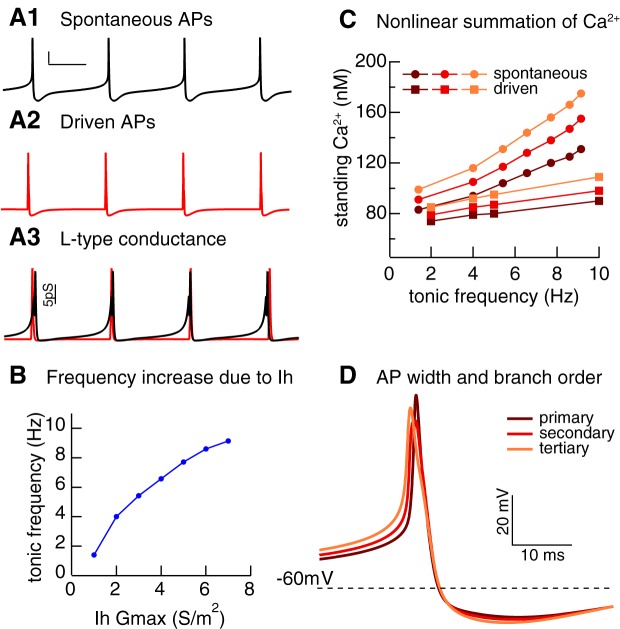
Fig. 3.
Ih alters standing calcium in model of substantia nigra pars compacta (SNc) dopamine neuron. A: spontaneous pacemaking, scale bars = 20 mV, 60 ms (A1). Action potentials (APs) evoked (A2) with a square depolarizing current pulse superimposed on a hyperpolarizing holding current to keep the neuron quiescent. A3: activation of L-type Ca2+ conductance in A1 (black) and A2 (red). B: tonic frequency increases with increased Ih conductance. C: standing calcium (defined as lowest calcium concentration reached between APs) increases more steeply with frequency in primary, secondary, and tertiary dendrites for pacemaking compared with driven APs as frequency is increased by stepping Ih Gmax from 1 to 7 S/m2. D: difference in AP width attributable to lower IA conductance density distally accounts for the difference in C. [Unpublished simulations by R. C. Evans.]

